Vertebrate Skeletal Anatomy I - The Skull
John Merck
Download cheat-sheet
Context I: Developmental regions
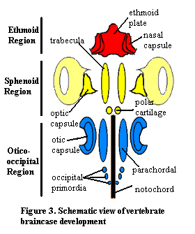
At the most basic level, the initial chondrifications (cartilage formation) of the neurocranium define the anteroposterior regionalization of the skull, with:
- Ethmoid region: Nasal capsules and supporting structures.
- Sphenoid region: Eyes and associated braincase structures derived from trabeculae - the primordial paired rods. In adults, this region supports the eyes and provides passage between the brain and olfactory capsules for the olfactory nerves.
- Otico-occipital region: ("Occipito-otic region" of some authors. Get used to seeing both terms.) Otic capsules and the brain, proper. Derived from primordial otic capsule, parachordal cartilages, and occipital arch cartilages.

Salvator merinae
- Between the sphenoid and otico-occipital region:
- The hypophysis (pituitary gland) occupies the recess where the trabeculae and parachordals meet. The small polar cartilages give rise to the pila antotica that form the back of the sella turcica (Turkish saddle) in which it sits. ("st" in linked image.)

C. Mimipiscis (actinopterygian), F Pucapapella (chondrichthyan) - The ventral cranial fissure separating ossifications of the braincase between the sphenoid and otico-occipital sections.

Mimipiscis (actinopterygian) - The hypophysis (pituitary gland) occupies the recess where the trabeculae and parachordals meet. The small polar cartilages give rise to the pila antotica that form the back of the sella turcica (Turkish saddle) in which it sits. ("st" in linked image.)
- Between the otic capsules and occipital arch: The lateral cranial fissure.
Chondrifications:
Major units of the skull are preformed in cartilage. Remember: all cartilages except for those of the otico-occipital region are preformed in migrating neural crest cells. The major units:
- The neurocranium: containing special sense capsules, brain, and supporting structures.(In blue - right)
- The mandibular arch: The paired elements of the jaws. (In yellow - right).
- Palatoquadrates: - upper jaws that articulate with the neurocranium dorsally.
- Meckel's cartilages: that articulate with the palatoquadrates.
- The hyoid arch: The paired elements supporting the jaws (although radically repurposed in land vertebrates!). (In green - right).
- Hyomandibulae: - that articulate with the neurocranium dorsally.
- Ceratohyals: that support the mandibular arch medially.
- Basihyals: Midline element to which both sides of the hyoid arch articulate.

Eusthenopteron foordi
In animals like the shark Clamydoselache these components and their relationships are clear. Among osteichthyans, we add a layer of dermal bones, but these are still associated with the major functional regions we've discussed.
In creatures like land vertebrates, they are not obvious, but they still provide the key to understanding cranial anatomy. Sources of obfuscation include:
- The integration of these functional units and blurring of their boundaries.
- The presence of superficial dermal elements of the skull.
- The fact that bony elements that ossify from them do not have a one-to-one correspondence with the cartilages from which they form.

Greererpeton burckemorani
Greererpeton:
We use the example of Greererpeton burckemorani, a creature near the common ancestry of land vertebrates. As such, it displays all of the cranial elements of Sarcopterygii for which homologies are securely known, but very few of them are high transformed from their ancestral state.
We continue to color-code:
- Blue: Ossifications of the neurocranium (dermal or endochondral)
- Yellow: Ossifications of the mandibular arch (dermal or endochondral)
- Green: Ossifications of the hyoid arch (endochondral)
- Pink: Ossifications of the skull roof (dermal)
- Orbits: Bony openings of eye sockets
- Nares (sing. Naris): Bony openings of nostrils
The palatal (ventral) aspect shows how ossifications of the skull roof, palatoquadrate, and neurocranium connect, but not too closely. Openings of the palate include:
- Choanae: (sing. Choana) Internal nostrils through which air enters oral chamber.
- Interpterygoid vacuity The space between the pterygoids (major ossifications of the palatoquadrate) on the midline
- Subtemporal fenestrae The "meat holes" through which the adductor muscles of the jaw pass.
Finally, the posterior view reveals:
- Posttemporal fenestrae Openings framed by the dermal skull roof and neurocranium.
In the course of vertebrate evolution, these openings are modified and, often, joined by new openings that we will need to keep track of. That's a story for later.


